Figure 4.
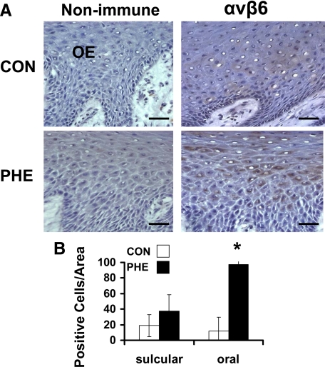
αvβ6 Integrin expression in phenytoin-induced gingival overgrowth and no overgrowth control tissues. A: Representative immunohistochemistry sections from oral epithelium (OE) of phenytoin (PHE)-induced gingival overgrowth and control (CON) tissues. Scale bar = 35 μm at ×400 magnification. B: Histomorphometric and quantitative analyses of αvβ6 integrin immunostaining in phenytoin-induced gingival overgrowth (PHE) in sulcular and oral epithelium compared with no overgrowth control (CON) tissues (0.09 mm2). Data are means ± SE; n = 5 for both control and phenytoin groups; *P < 0.05 compared with control by analysis of variance with Bonferroni correction for multiple tests.

