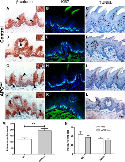
Figure 1.
Increased nuclear β-catenin in the tongue epithelium of APCmin/+ mice. A, D, G, and J: The tongue epithelium of APCmin/+ mice (G and J) shows increased nuclear localization of β-catenin compared with controls (A and D). Note that positive nuclei are located in the epithelium and in taste buds; arrowheads point to nuclear β-catenin. M: Quantification of β-catenin positive nuclei in APCmin/+ compared with wild-type (WT) mice (**P < 0.005). B, E, H, and K: Cell proliferation was not altered in APCmin/+ mice (H and K) compared with controls (B and E), determined by Ki-67 antigen immunofluorescence. C, F, I, and L: apoptosis was not significantly altered in the tongue epithelium of APCmin/+ mice (I and L) compared with wild-type (C and F) as evaluated by TUNEL staining. N: Combined graph showing quantification of Ki-67 and TUNEL positive cells in APCmin/+ mice compared with wild-type. For statistical analysis, three animals of each genotype were analyzed for nuclear β-catenin, Ki-67, and TUNEL staining.

