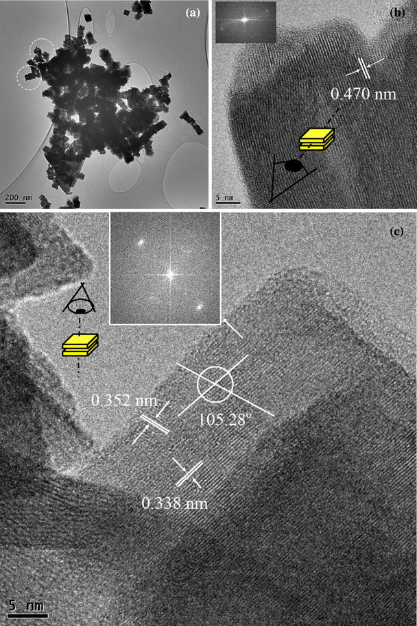
Figure 3.

a Low magnification TEM image of the WOx showing stacking of nano-platelets, b cross-sectional view of one stacking, showing each platelet can be up to 20 nm thick with one preferred direction of stacking as shown by the inset of the image’s FFT. c Top-view of two platelets stacked together showing the inter-planar spacing, angles and the two-dimensional growth shown by FFT of the image in the inset of c
