
Figure 3.
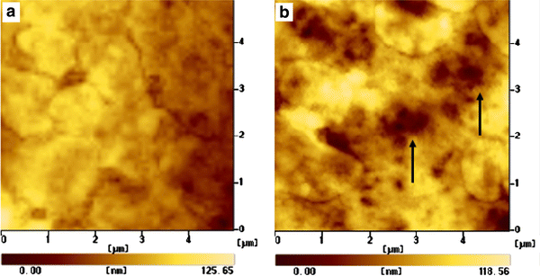
AFM images for cell uptake of Ni nanoparticles.aSMMC-7721 cell without Ni nanoparticles (control experiment); andbthe cell surface after incubation with 12.5 μg/mL of Ni nanoparticles for 6 h, which illustrates the apparent uptake of Ni nanoparticles during the process of endocytosis that led to the change of the cell membrane and formation of holes (arrows) on the surface of the relevant cell