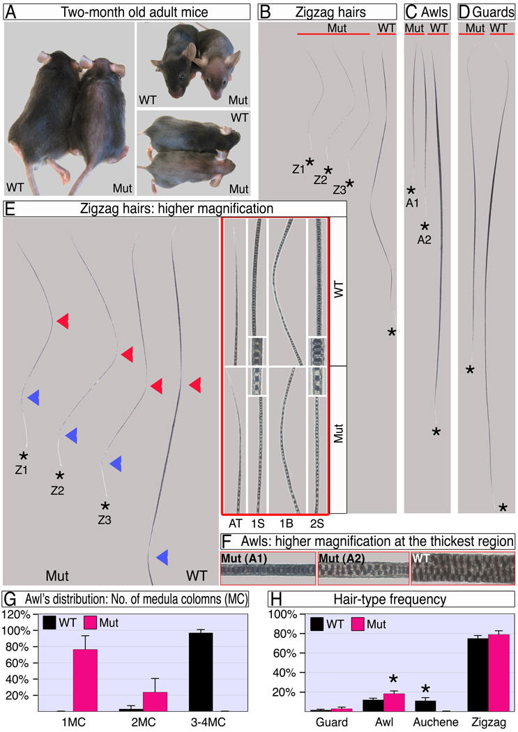
Figure 2. Ablation of β-catenin in the DP results in dramatic hair shortening and thinning.
(A) Adult wild type and mutant mice. Note the epidermis is visible through the thin mutant hair coat. (B–D) Zigzag, awl and guard hair-types after the first hair cycle. The apical tips of all hairs are aligned next to the red line and hair-clubs are marked by stars. Mutant hairs within a hair-type population are classified into sub-types based on their different length and/or thickness. Z1–Z3 in B represent a first segment 50% or less, between 50–100%, and similar in length to wild type respectively. A1–A2 in C represent 1 or 2 columns of medulla cells respectively. (E) Mutant zigzag hairs and the apical portion of a wild type zigzag are shown at left. The first and second bends from the apical tip are marked by red and blue arrowheads, respectively. At right, framed with red line, higher magnifications of the apical tip (AT), mid-domain of the first segment (1S), first bend (1B) and mid-region of the second segment (2S) reveal the reduced thickness in mutant hair along the entire hair shaft. Insets show the thickness and organization of the medulla column. (F) Higher magnifications of the thickest region of awl hairs show the number, structure and organization of the medulla columns. (G) Distribution of awl hairs according to the number of medulla columns (MC) at the thickest region. (H) Frequency of hair types in wild type and mutant. Two-tailed unpaired students T-test was employed (*, p<0.0001). See also Figure S1.

