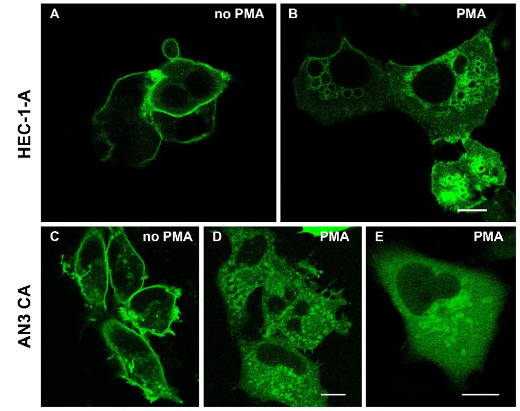
Figure 5.
Fluorescent images of HEC-1-A (A, B) and AN3 CA cells (C, D, E) transfected with a full-length gravin-EGFP construct. The gravin-EGFP fusion protein localized at the cell periphery in untreated cells (A, C), but translocated to juxtanuclear vesicles after PMA treatment (B, D, E). All scale bars, 10 µm.

