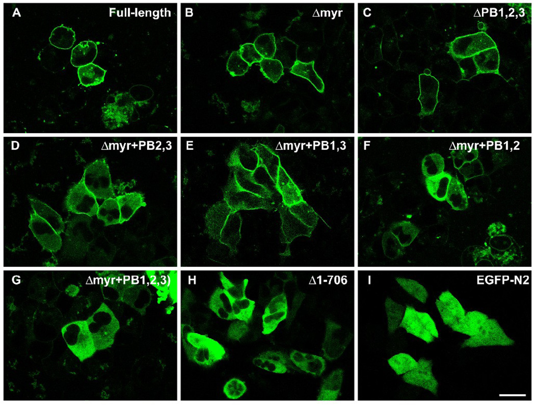
Figure 7.
Confocal images of AN3 CA cells expressing full-length gravin-EGFP (A), mutant gravin-EGFP constructs lacking different portions of the proposed membrane targeting domains as labeled (B–H), and EGFP-N2 alone (I). Deletion of the myristoylation site or all three polybasic regions did not significantly change the peripheral distribution of gravin-EGFP fusion protein (B, C). Mutant gravin-EGFP fusion proteins lacking the myristoylation site and different combinations of any two of the three polybasic regions lost the peripheral distribution to some degree (D–F). Deletion of the myristoylation site and all three polybasic region resulted in a construct that displayed a strictly cytosolic distribution similar to that displayed by EGFP-N2 except for the absence of nuclear labeling (G–I). Scale bar, 20 µm.

