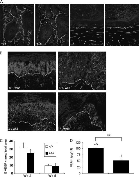
Fig. 6.
MMP13 mediates the release of VEGF from the ECM. (A) Immunostaining on parallel cryosections of s.c. tumours from Mmp13+/+ (panels 1 and 2) and Mmp13−/− mice (panels 3 and 4) for VEGF (red, panels 1 and 3), VEGFR-2 (red; panels 2 and 4), CD31 (green) and pan-keratin (blue), bar 100 μm. (B) Non-radioactive in situ hybridization with a murine VEGF-164 probe (red) of early (left panels) and late (right panels) surface transplants of Mmp13+/+ (upper panels) and Mmp13−/− (lower panels) mice, with immunofluorescent staining of blood vessels against collagen IV (green) and tumour tissue against pan-keratin (blue), bar 200 μm. The line marks the tumour stroma border. (C) Quantification of VEGF expression in surface transplants of Mmp13+/+ and Mmp13−/− mice (VEGF-positive area/total area). (D) Release of human VEGF-165 from a collagen type I gel containing Mmp13+/+ or Mmp13−/− MEFs, VEGF levels were measured by enzyme-linked immunosorbent assay, data shown for conditioned media of day 6 with 500 pg/ml of VEGF in the gel, **P < 0.001.

