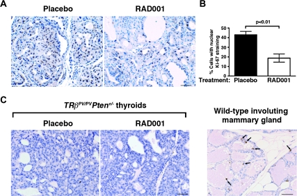
Fig. 2.
Inhibition of mTORC1 signaling reduces cell proliferation without affecting cell apoptosis in FTC. (A) Representative microphotographs of Ki-67 immunohistochemistry on sections of placebo- and RAD001-treated TRβPV/PVPten+/− thyroids counterstained by hematoxylin; bar, 50 μm. (B) Thyroid cell proliferative index, determined by Ki-67 immunohistochemistry in the two groups, shows that there is a significant reduction in the percentage of proliferating cells in the thyroids of RAD001-treated TRβPV/PVPten+/− mice (n = 4 mice) as compared with placebo-treated TRβPV/PVPten+/− mice (n = 5 mice). (C) Representative microphotographs showing no increase in apoptosis in RAD001-treated animals after prolonged treatment, as measured by terminal deoxynucleotidyl transferase-mediated dUTP nick end labeling assay. Involuting wild-type female mammary gland, used as a positive control for terminal deoxynucleotidyl transferase-mediated dUTP nick end labeling assay, shows apoptotic epithelial cells (depicted by arrows); bar, 50 μm.

