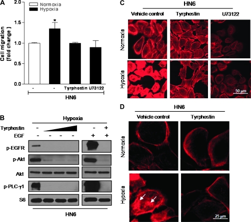
Fig. 4.
Inhibition of EGFR/PLC-γ1 activation blocks actin redistribution and cell migration. (A) Pretreatment with the EGFR tyrosine kinase inhibitor tyrphostin (25 μM) or U73122 (3 μM) inhibited the increased migratory capacity of hypoxic cells. (B) HN6 cells were exposed to hypoxia for 6 h in the presence or absence of increasing concentrations of tyrphostin (5–50 μM). Western blotting evaluated phosphorylated status of EGFR, Akt and PLC-γ1. EGF (100 ng/ml) was used as a positive control for EGFR-mediated signaling. S6 was used as sample loading control. (C) Actin cytoskeletal changes were prevented following pretreatment with tyrphostin (25 μM) or U73122 (3 μM) prior to 6-h exposure to hypoxia. (D) Higher magnification image depicts redistribution of actin aggregates in hypoxic cells (arrows) when compared with normoxic controls or cells pretreated with tyrphostin (25 μM).

