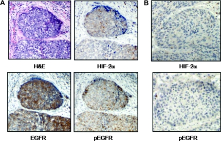
Fig. 6.
EGFR phosphorylation within hypoxic tumor foci in human HNSCC. (A) Representative human HNSCC specimen where immunohistochemistry was performed using antibodies against HIF-2α, total EGFR and phospho-EGFR along with hematoxylin and eosin (H&E). Phospho-EGFR staining was detected within hypoxic areas as judged by HIF-2α accumulation. (B) Another representative HNSCC specimen in which HIF-2α immunostaining was negative, undetectable levels of phospho-EGFR were found. Magnification, ×100.

