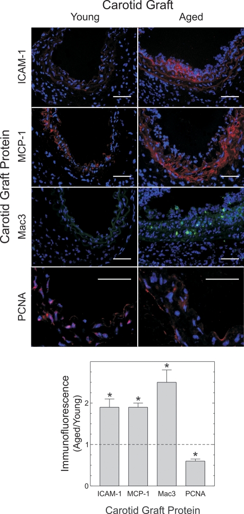
Figure 3.
Aged arteries demonstrate greater macrophage recruitment and less SMC proliferation than young arteries during early atherogenesis. Two-week-old grafts from Figure 2 were immunostained for the indicated proteins and counterstained for nuclear DNA (blue). Protein immunofluorescence was then normalized to nuclear fluorescence within each microscopic field, as described in Materials and Methods. The means ± SE from four or more arteries are plotted. Compared with young specimens: *P < 0.05. Scale bars: 50 μm (original magnification ×440).

