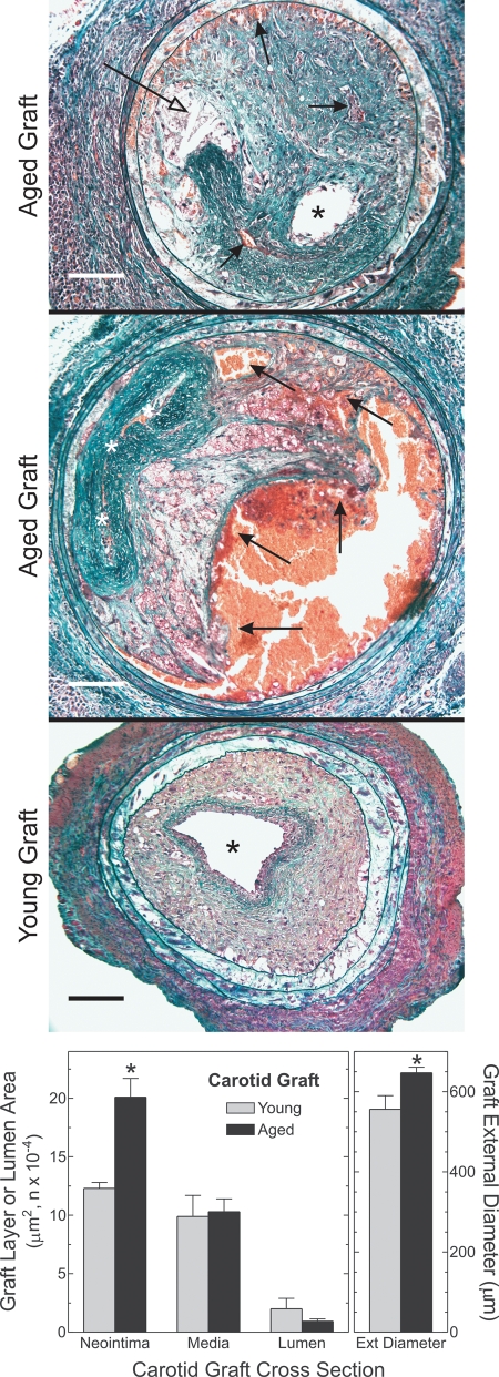
Figure 4.
Arterial wall aging exacerbates plaque hemorrhage and arterial outward remodeling. Carotid grafts were harvested 7 weeks post-operatively, after perfusion–fixation. Paraffin sections were stained with a modified connective tissue stain (see Materials and Methods). Computerized morphometry yielded the indicated dimensions from six or more arteries of each type. Compared with young grafts: *P < 0.01. Open arrows, closed arrows and asterisks indicate cholesterol clefts, plaque hemorrhage and lumen, respectively. Scale bars: 100 μm (original magnification ×200). The middle image was concatenated with Image-Pro Plus® (Media Cybernetics).

