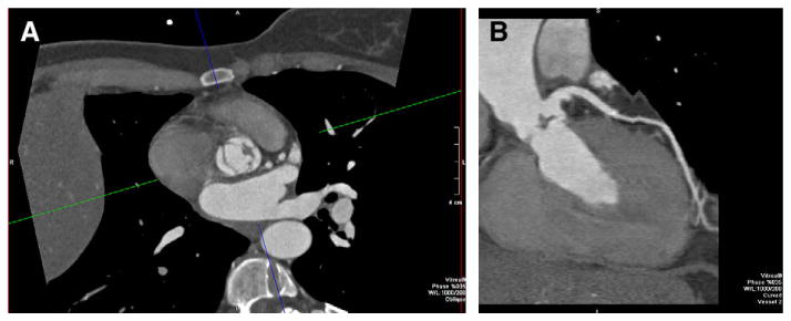
Fig. 3.

55-year-old man with abnormal aortic valve motion by echocardiography. CT was requested to exclude CAD. The patient's heart rate (>100 beats per minute at the time of acquisition) could not be safely lowered because of hypotension. (A) Retrospectively ECG gated dual-source cardiac CT (Siemens Definition, Erlangen, Germany) with 83 milliseconds temporal resolution depicted the vegetation of the aortic valve, as noted on the axial images. (B) Curved multiplanar reformatted image of the LAD also shows the vegetation in addition to demonstrating a normal LAD.
