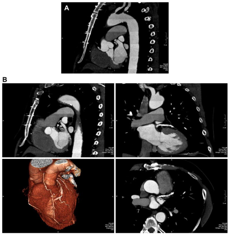
Fig. 4.

72-year-old man status post coronary artery bypass grafting. Because of the large craniocaudal field of view, imaging was performed over 13 heartbeats, 7 beats of data acquisition plus 6 move the patient within the gantry. This “step and shoot” technique significantly decreases the radiation exposure when compared to retrospective ECG gating. (A) Left anterior oblique reformation over the entire z-axis FOV shows high image quality. Enhancement pattern in the aorta demonstrates the individual “steps” as described. (B) Multiplanar reformatted images as well as 3D volume rendering (lower left) focused on the heavily calcified left anterior descending.
