
Fig. 1.
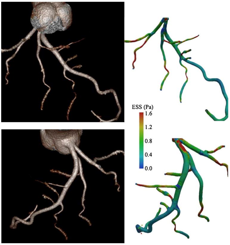
Whole intracoronary endothelial shear stress (ESS) mapping from single-heartbeat 320-detector row CT (Toshiba Aquilion One Dynamic Volume CT; Tochigi, Japan) after injection of 80 mL of iopamidol, 370 mg I/mL (Isovue-370; Bracco Diagnostics, Princeton, NJ), followed by 40 mL of normal saline injected with a dual injector (EZEM Empower CTA DUAL Injector; EZEM Inc., Lake Success, NY). Three-dimensional volume-rendered images generated from Vitrea 4.1 software (Vital Images, Minnetonka, MN) (left side). Corresponding ESS maps (right side). Simulated blood flow uses a computational fluid dynamic technique. (Adapted from Ramkumar et al. [59]; with permission)