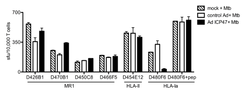
Figure 3. MR1-restricted recognition of Mtb-infected cells is TAP-independent.
DCs autologous to D454 and expressing HLA-B08 were transduced with either a control adenoviral vector or adenoviral ICP47 using lipofectamine 2000. After 16 h, DCs were washed and either left uninfected, infected with Mtb, or pulsed with HLA-B08 specific peptide CFP103–11. Following overnight incubation, T cells were added (10,000) to DCs (25,000/well) and IFN-γ production was assessed by ELISPOT. Results are representative of three independent assays. No responses were detected from T cells incubated with uninfected DCs with or without adenoviral vectors. Error bars represent the mean and standard error from duplicate wells.

