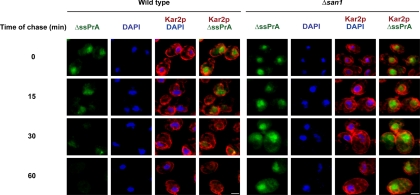
Figure 5.
Visualization of intracellular substrate decay. Logarithmically growing wild-type and Δsan1 cells expressing ΔssPrA were treated with cycloheximide (200 μg/ml final) to terminate translation. At times indicated, cell aliquots were taken, fixed, and stained for ΔssPrA and Kar2p as described in Figure 2. Image acquisition times and settings were identical for each series. Scale bars, 2 μm.

