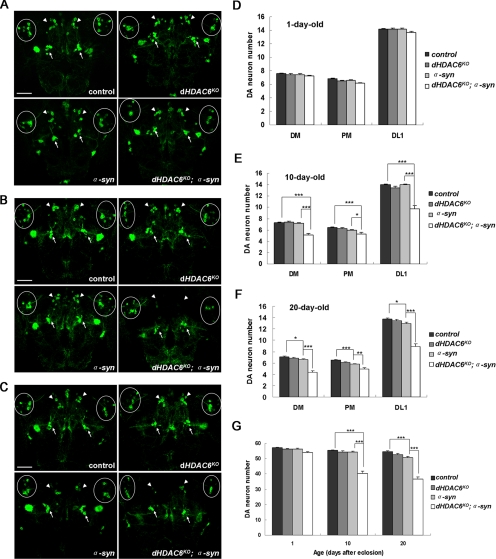
Figure 2.
dHDAC6KO promotes α-synuclein–induced dopaminergic neuron loss. Confocal images of dopaminergic neurons in DM, PM, and DL1 regions immunostained with anti-GFP antibody on 1-d-old (A), 10-d-old (B), and 20-d-old (C) fly brains, respectively, are shown, with arrows for PM and arrowheads for DM clusters and circles for DL1 clusters. (D–F) Quantitative graphs are shown corresponding to confocal images of 1-d-old, 10-d-old, and 20-d-old fly brains, respectively. (G) Graphs showing total numbers of dopaminergic neurons in the DM, PM, and DL1 clusters of different genotypes as indicated at 1 d, 10 d, 20 d. Data were analyzed by Student's t test and presented as mean ± SEM (n = 20∼40) with * for p < 0.05, ** for p < 0.01, and *** for p < 0.001. Bar, 50 μm. Genotypes: control flies are w; UAS-mCD8::GFP/+; TH-GAL4/+. dHDAC6KO flies are w, dHDAC6KO; UAS-mCD8::GFP/+; TH-GAL4/+. α-syn flies are w; UAS-mCD8::GFP/+; TH-GAL4, UAS-α-synuclein/+. dHDAC6KO; α-syn flies are w, dHDAC6KO; UAS-mCD8::GFP/+; TH-GAL4, UAS-α-synuclein/+.

