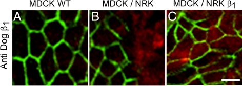
Figure 1.
MDCK cells express β1 subunits at homotypic but not at heterotypic contacts. Localization of the dog β1 subunit (A–C, green) of Na+,K+-ATPase was studied by immunofluorescence assay in MDCK cells derived from dog kidney. (A) Monolayer of pure MDCK cells showing that the β1 subunit is only expressed at the plasma membranes in the lateral domain where cells contact each other. (B) MDCK cells cocultured with NRK cells (derived from normal rat kidney) that were labeled beforehand with CMTMR (red). In the mixed monolayer, the β1 subunit is only expressed at homotypic borders (MDCK/MDCK) but not at heterotypic ones (MDCK/NRK). (C) Monolayer of mixed MDCK/NRK cells transfected with dog β1 subunit shows that the β subunit (green) is concentrated at both homotypic MDCK/MDCK contacts and heterotypic MDCK/NRK contacts. Bars, 20 μm.

