Figure 6.
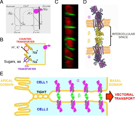
Polarized expression of epithelial Na+,K+-ATPase. (A) Seminal model of Koefoed-Johnsen and Ussing (1958), in which the Na+,K+-ATPase (reinforced in magenta) was assumed to occupy the basal side of the cell, which constitutes the inner-facing barrier. (B) From this position, the pump transports Na+ toward the interstitial side of the cell, producing a net decrease in the cytoplasmic concentration of this ion and setting up an electrochemical force that drives counter- and cotransporters of sugars, amino acids, and ions and possible the existence of metazoans. (C) Confocal transverse section of a monolayer of MDCK cells. The nuclei are stained with propidium iodide (red) and the β subunits of Na+,K+-ATPase are stained with a specific antibody (green), showing that this subunit is localized to the lateral surfaces of cells, but not to the apical (left) or basal sides. (D) Na+,K+-ATPase molecules of two neighboring epithelial cells with interacting β subunits (green), as shown previously (Shoshani et al., 2005) and in the present work. (E) Na+,K+-ATPase molecules anchored to the lateral membranes. Because of the tight junction, Na+ ions pumped into the intercellular space can only diffuse inwards, generating vectorial transport across the epithelium.

