Figure 2.
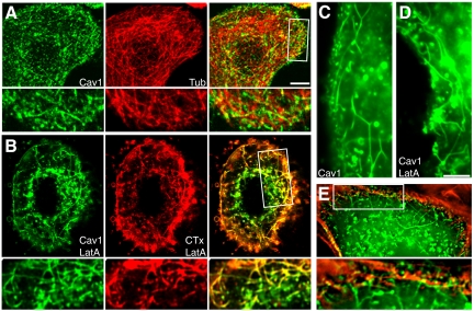
Interactions of caveolin-1 tubules with actin and microtubule cytoskeletons in SK-BR-3 cells. (A) Cells expressing caveolin-1-GFP (left) and mCherry-α-tubulin (middle) were visualized by fluorescence microscopy. Caveolin-1-GFP tubules rarely colocalized with microtubules. (A, B, and E) High-magnification views of the boxed region in the merged images are shown beneath each panel. (B) Cells expressing caveolin-1-GFP (left) were treated with 5 μM LatA for 30 min. AF-594-CTxB (middle) was added for the last 10 min, and cells were fixed and examined by fluorescence microscopy. (C and D) Morphology of tubules in caveolin-1-GFP–expressing control (C) and LatA-treated (D) cells. (E) A cell expressing caveolin-1-GFP was fixed, permeabilized, and incubated with AF-594-phalloidin to label actin filaments. Short tubules and puncta associated with actin filaments under the plasma membrane, whereas long tubules extended into the cell from the actin-rich subcortical region. An enlarged view of the boxed region is shown beneath the panel. Bars, 5 μm (bar in A applies to A and B; bar in D applies to C-E).

