Figure 3.
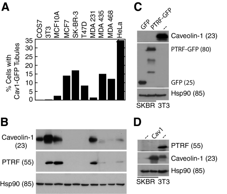
Caveolin-1-GFP tubules correlate with loss of endogenous caveolin-1 and PTRF/cavin-1; forced expression of PTRF/cavin-1 or caveolin-1 does not induce endogenous expression of the other gene in SK-BR-3 cells. (A) Caveolin-1-GFP was expressed in COS7, NIH 3T3, MCF-10A, MCF-7, SK-BR-3, T47D, MDA-MB-231, MDA-MB-435, MDA-MB-468, and HeLa cells. The percentage of transfected cells in which caveolin-1 was detected in tubules by fluorescence microscopy is indicated (average of at least 2 experiments that varied by <15%, counting at least 150 transfected cells in each). (B) Lysates of the cells listed in A were analyzed by SDS-PAGE and Western blotting, detecting caveolin-1, PTRF/cavin-1, and Hsp90 as a loading control. Cell line labels in A also refer to the corresponding bands in B. (C) SK-BR-3 cells (first 2 lanes) were transfected with GFP or PTRF/cavin-1-GFP as indicated (top of blot) and grown for 3 d before harvest. Lysates of these cells and of untransfected NIH 3T3 cells (third lane) containing 25 μg of protein were subjected to SDS-PAGE and transfer to polyvinylidene difluoride (PVDF). The blot was probed sequentially with anti-caveolin-1, anti-GFP, and anti-Hsp90, stripping between probings. (D) SK-BR-3 cells (first 2 lanes) were infected with caveolin-1-expressing adenovirus as indicated (top of blot), or left uninfected, and grown for 4 d before harvest. Lysates of these cells and of uninfected NIH 3T3 cells (third lane) containing 25 μg of protein were subjected to SDS-PAGE and transfer to PVDF. The blot was probed sequentially with anti-PTRF/cavin-1, anti-caveolin-1, and anti-Hsp90, stripping between probings. (B–D) Molecular masses (in parentheses) are in kilodaltons.

