Figure 5.
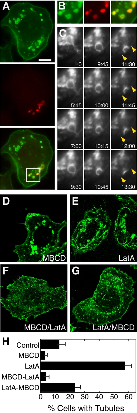
MBCD alters caveolin-1-GFP localization and reduces tubule frequency in SK-BR-3 cells. (A) SK-BR-3 cells expressing caveolin-1 GFP (top) were incubated with 10 mM MBCD for 45 min and then with 1 mM fluoro-ruby red dextran (middle) for 5 min before fixation and detection by fluorescence microscopy. (B) Enlarged views of the boxed region of the merged image of the cell shown in A. (C) Frames from Supplemental Movie 2, showing a live MBCD-treated SK-BR-3 cell expressing caveolin-1-GFP. A caveolin-1-GFP–positive vacuole that initially seems tethered to the plasma membrane approaches the plasma membrane (indicated with arrows in the last four frames) and fuses with it. Time elapsed (minutes:seconds) after the first frame is shown. (D–G) Images of caveolin-1-GFP–expressing SK-BR-3 cells treated with MBCD (30 min; D), LatA (10 min; E), MBCD (30 min) and then MBCD + LatA (10 min; F), or LatA (10 min) and then MBCD + LatA (30 min; G). Bar, 10 μm. (H) Frequency of caveolin-1 long tubules in cells treated as in D–G. The mean ± SEM of at least three experiments (counting at least 150 cells on each slide) is shown.

