Figure 8.
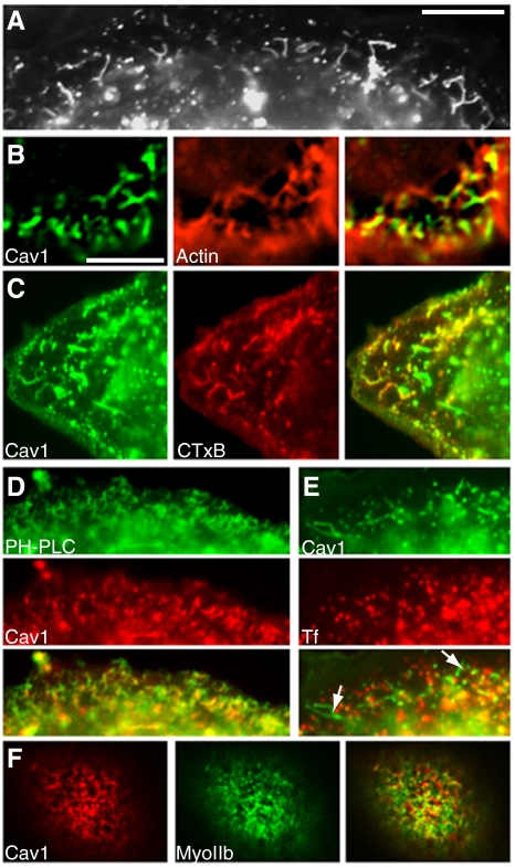
Short caveolin-1 tubules in SK-BR-3 cells are linked to actin and derived from the plasma membrane. (A) Section of a caveolin-1-GFP–expressing cell shows short, branched tubules just under the cell surface. (B) Cells expressing caveolin-1-GFP (left) were fixed, permeabilized, and treated with AF-594-phalloidin (middle). A cell section just under the plasma membrane (at the bottom and right of the image), containing short tubules close to actin filaments, is shown. (C) Cells expressing caveolin-1-GFP (left) were incubated with AF-594 CTxB (middle) for 2 min before fixation. Internalized CTxB-labeled puncta and short tubules near the cell periphery. (D) Cells coexpressing GFP-PH-PLCδ (top) and caveolin-1-RFP (middle) were processed for fluorescence microscopy. Caveolin-1-RFP–positive short tubules and puncta are labeled with GFP-PH-PLCδ. (E) Cells expressing caveolin-1-GFP (top) were incubated with AF-594 transferrin (middle) for 5 min. Caveolin-1-GFP–positive short tubules (arrows in merged image; bottom) are not labeled with transferrin. (F) Cells coexpressing caveolin-1-RFP (left) and GFP-myosin IIa (middle) were fixed and examined by fluorescence microscopy. High-magnification views of the boxed region in the merged image (right) are shown beneath each panel. Additional images of the same cell, showing dorsal stress fibers below the zone containing short tubules, are shown in Supplemental Figure 6. Bar in A, 10 μm. Bar in B (applies to B–F), 5 μm.

