
Figure 4.
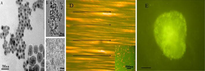
TEM images of a FMCNPs b MNPs and c quantum dots. d FMCNPs are aligned in a magnetic field obtained with a fluorescent microscope3. The arrows are added to clearly show the orientation of the magnetic field. The inset image is obtained without a magnetic filed. e Fluorescent microscope image of FMCNPs inside murine ECC stem cells, scale bar: 10 μm [53]