Figure 5.
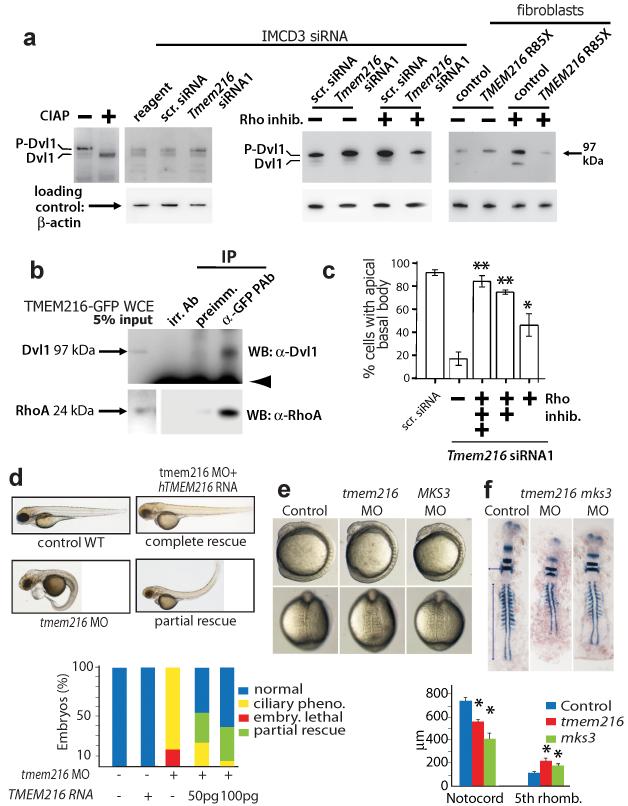
TMEM216 disruption results in Dvl1 phosphorylation, and planar cell polarity-like phenotypes in zebrafish. (a) siRNA knockdown of Tmem216 (left panel) and TMEM216 p.R85X patient fibroblasts (right panel) cause an increase in the upper (phosphorylated) isoform (P-Dvl1) compared to scrambled control (scr.). Treatment with Rho inhibitor exoenzyme-C3-transferase (2 μg/ml) alone increased Dvl1 phosphorylation, but increases in P-Dvl1 by TMEM216 loss are reversed by Rho inhibition (right panel). (b) Co-immunoprecipitation of both Dvl1 and RhoA with TMEM216 in TMEM216-GFP transfected cells. Arrowhead is IgG heavy chain. (c) Dose-dependent rescue of centrosome/basal body docking phenotype in Tmem216 siRNA-treated cells following += 0.5, ++= 1.0, +++= 2.0 μg/ml Rho inhibitor treatment. *p<0.01; **p<0.001 for chi-squared test. (d) Injection of translation-blocking morpholino (MO) to tmem216 vs. scrambled MO causes a ciliary defect phenotype in injected zebrafish embryos (>50 each condition). Injection of human TMEM216 RNA causes no phenotype in WT embryos, but allows partial, dose-dependent rescue of the MO phenotype. (e) Lateral (top) and dorsal (bottom) views of zebrafish embryos injected with tmem216 or mks3 MO at 8-somite stage had ciliopathy features. (f) Representative 11-somite stage embryos hybridized with krox20, pax2, and myoD riboprobes. Convergence to the midline is measured by the width at the fifth rhombomere (horizontal arrow), and extension along the anterior-posterior (AP) axis by notochord length (vertical arrow) (n=12-15 embryos/injection). Suppression of the tmem216 or mks3 morphant gastrulation defect causes significant differences in width and length compared to controls (*p<0.005). Pheno.=phenotype; embry.=embryonic; rhomb.=rhombomere; Bars=standard error of means.

