
Figure 2.
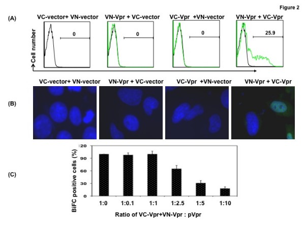
Visualization of Vpr oligomerization by (A) Flow cytometry or (B) Fluorescent microscopy. (A) Quantitative analysis by flow cytometry of Venus fragment complementation in HeLa cells transfected with VC-Vpr and VN-Vpr or with control plasmid. Thirty-six hours posttransfection, cells were harvested and analyzed by flow cytometry to determine the percentage of cells positive for BiFC fluorescence. Results represent the means of five independent experiments. (B) Subcellular localization of the BiFC complex: HeLa cells grown on glass coverslips were cotransfected with VC and VN plasmids, VN-Vprwt and VC-Vprwt or VN- Vprwt or VC- Vprwt with control plasmid pairs using Lipofectamine. At 36 hours post-transfection, cells were fixed, stained with DAPI and imaged at 60× magnification. (C) To confirm the specificity of VC and VN based oligomerization, VC-Vpr (0.5 ug) and VN-Vpr (0.5 ug) plasmids were cotransfected with increasing concentrations of empty vector or Vpr expression plasmid. Thirty-six hours post transfection cells were fixed and assessed by flow cytometry. BiFC positive cells (%) were calculated in each treatment compared with the control. Figure represents one of five independent experiments (n = 5) with similar results.