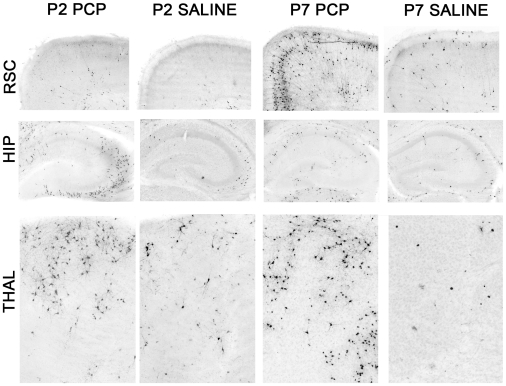
Figure 1. Pattern of neuroapoptosis following PCP treatment in neonatal mice differs depending on whether exposure occurs on P2 or P7.
Photomicrographs showing activated caspase3- (AC3-) positive cell profiles indicating neuroapoptotic damage in the retrosplenial cortex (RSC), hippocampus (HIP), OR anterodorsal thalamic nuclei (THAL) in mice that were treated with PCP on either P2 (P2PCP) or P7 (P7PCP) or with saline on the same postnatal days (P2 Saline and P7 Saline, respectively). PCP exposure on P2 produced neuroapoptotic damage to the HIP and THAL regions without affecting the RSC. In contrast, PCP exposure on P7 resulted in severe neuroapoptosis in the RSC and moderate damage to the THAL, while leaving the HIP relatively spared. Images taken at 10× magnification.

