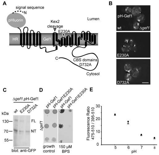
Fig. 4.
Methodology to determine the pH gradient across the Gef1-containing compartment. (A) Scheme of the Gef1 fusion protein used as a probe to assess pH inside the Gef1-containing compartment. The signal sequence of invertase aids translocation of the pH-sensitive GFP pHluorin to the lumen. The archeal membrane protein halorhodopsin serves to adapt the luminal pHluorin to the cytosolic N-terminus of Gef1. All other details as indicated in Fig. 1A. (B) Subcellular localisation of N-terminal pHluorin-HR-Gef1 fusions with the indicated mutations revealed by live-cell imaging in a Δgef1 strain. Scale bar: 5 μm (C) Anti-GFP western blot of total cellular lysates from cells expressing the indicated pHluorin-HR-Gef1 fusion proteins. ‘FL’ and ‘NT’ indicate the migration position of uncleaved full-length Gef1 and the N-terminal product of the cleavage reaction, respectively. (D) The fusion protein used as a luminal pH probe complements the growth defect of a Δgef1 strain on low-iron medium. Serial dilutions of cultures grown in SC medium were spotted on a SC plate without (‘growth control’) or with 150 μM of the iron chelator BPS as indicated. (E) Calibration curves of the cytosolic (triangles) and luminal (circles) pHluorin probes. Background-corrected fluorescence ratios of both probes are reported as a function of pH after digitonin treatment in the indicated buffer.

