
Fig. 7.
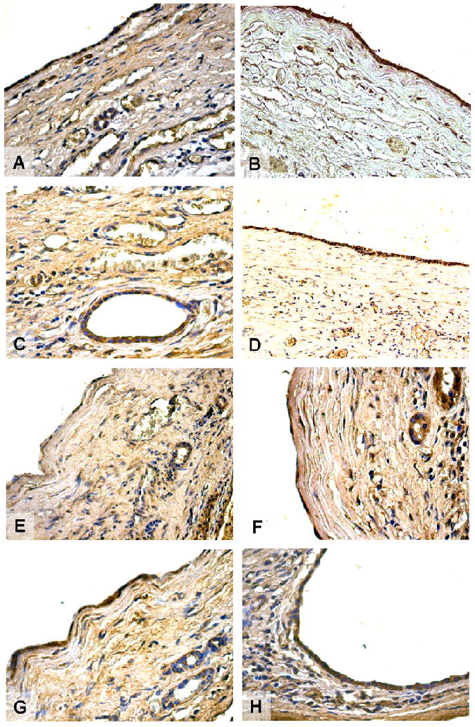
Immunohistochemistry for ER-α (A), ER-β (B), IGF-1 (C), IGF1-R (D), FSH (E), FSHR (F), VEGF-A (G), and VEGF-C (H) in hepatic cysts from patients with ADPCLD. Cholangiocytes of reactive bile ducts close to the cysts are positive for both estrogen receptors. The epithelium lining small and large cysts showed strong positivity for ER-α and ER-β located at cytoplasmic levels. Both IGF1 and its receptor showed positive immunolocalisation in biliary epithelium. The expression of the hormone FSH is also present in both small and large cysts. A stronger immunolocalisation is found for FSHR in the biliary epithelium that lines hepatic cysts. Furthermore, biliary epithelium of small and large cysts shows an high positivity for VEGF-A and VEGF-C, that play an important role in the growth of cysts and in the progression of PCLD. Original magnification 20 ×.