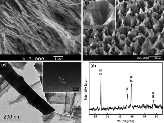
Figure 1.

a SEM image of Bi nanowires embedded in AMM and grown out of the membranes. b SEM image of the bunch-like Bi composed of Bi nanowires. The inset shows the nest formed by the nanowires. c TEM image of individual Bi nanowires embedded in the AAM. The inset shows the corresponding lattice fringe image. d Typical XRD pattern of the prepared bunch-like Bi electrode
