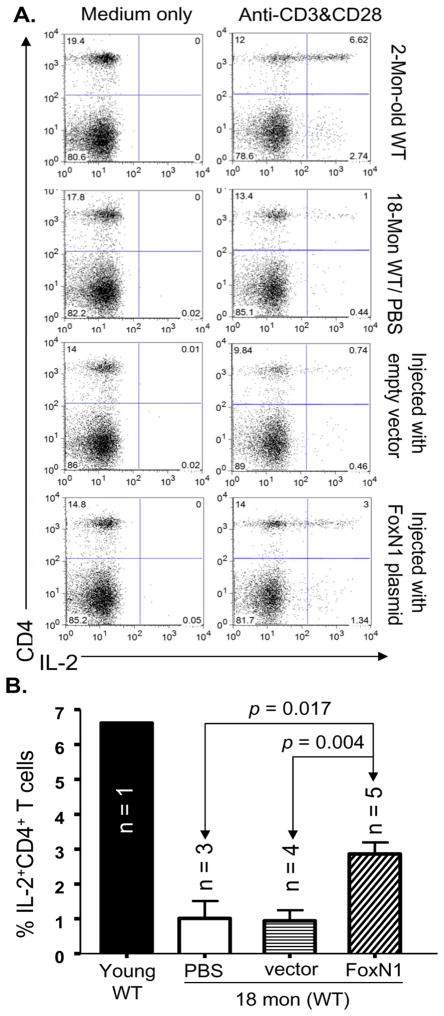
Figure 8. Increased IL-2 production in peripheral CD4+ T cells of aged mice via intrathymic infusion of exogenous FoxN1-cDNA.
(A) A representative FACS result, from top to bottom, shows IL-2+CD4+T-cell subset in young mouse spleen (2 months old), aged mouse spleen (18 months old), empty vector-infused aged mouse spleen (18 months old), and FoxN1-cDNA-infused aged mouse spleen (18 months old) without stimulation (left panels) and in response to CD3 and CD28 antibodies (right panels). (B) A summarized % of splenic IL-2+CD4+T cells of the above four groups in response to CD3 and CD28 antibodies.

