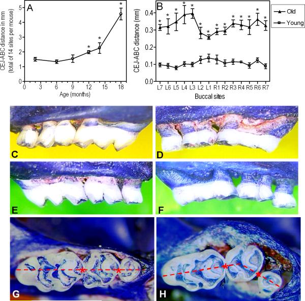
Fig. 1.
Periodontal bone loss as a function of age in BALB/c mice. (A): Young (8-10 weeks of age), old (≥ 18 months of age), and mice of intermediate ages (6-, 9-, 12-, and 14-month-old) were used to determine their periodontal bone levels. The mm distance from the cementoenamel junction (CEJ) to the alveolar bone crest (ABC) was measured at 14 predetermined maxillary buccal sites and the readings were totaled for each mouse. The data are means ± SD (n = 5 mice). (B): Analytical data from the two extreme age groups (young vs. old). Each point corresponds to a measured site (L1-L7, left maxilla; R1-R7, right maxilla) and represents means ± SD (n = 5 mice). Asterisks denote significant (p < 0.05) differences in CEJ-ABC distances compared to young mice (the greater the CEJ-ABC distance, the greater the bone loss). The experiment was repeated with additional sets of 5 mice per group yielding consistent results. (C-F): Representative images from the maxillae of young (C, right; E, left) and old (D, right; F, left) mice. Extensive areas of resorbed alveolar bone are evident in the old mice. (G-H): Maxillary molar blocks seen from the occlusal surfaces of young (G) and old (H) mice. Note that all three molars in young mice are in line with each other (i.e., a straight line can connect their contact points, indicated by asterisks). Due to tooth mobility, this relationship did not apply to the molar blocks of old mice, many of which displayed overt migration of molars (especially of the second molar in buccal direction).

