
Figure 5.
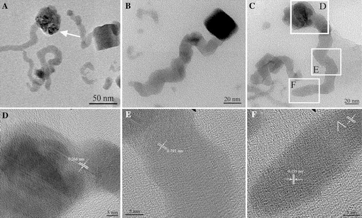
TEM images showing oriented attachment of copper selenide nanosnakes in BSA solution for 48 h. a Low-magnification TEM image of sample. b, c TEM images of two different devour stages of copper selenide nanosnakes. HRTEM images of different parts of an individual nanosnake: d the neck, e the body, f the tail, respectively