Figure 4.
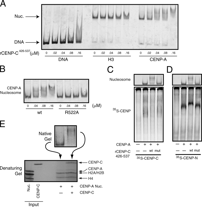
CENP-C and CENP-N bind to different sites on the same CENP-A nucleosomes. (A) α-Satellite DNA or reconstituted nucleosomes (10 nM) were incubated with the indicated concentration of rCENP-C426–537 and resolved on native gels. DNA or nucleosomes were visualized after staining with SYBR gold. (B) CENP-A nucleosomes (10 nM) were incubated with the indicated concentration of wild-type (wt) or mutant (R522A) rCENP-C426–537 and resolved on a native gel. (C) [35S]methionine-labeled CENP-C was incubated alone (−) or in the presence of 150 nM of CENP-A nucleosomes (+). In addition, binding reactions contained wild-type (wt) or the R522A mutant (mut) rCENP-C426–537 (300 nM), or buffer alone (−). The gel was stained with ethidium bromide to visualize nucleosomes (top) and was subsequently dried and scanned on a phosphorimager to visualize the labeled CENP-C (bottom). (D) An identical experiment to C except that [35S]methionine-labeled CENP-N was used. (E) Isolated nuclei were digested with micrococcal nuclease, and wild-type or R11A mutant GFP–CENP-N was immunoprecipitated using anti-GFP antibodies. Each immunoprecipitate was probed with the indicated antibodies. Control cells don’t express GFP–CENP-N.

