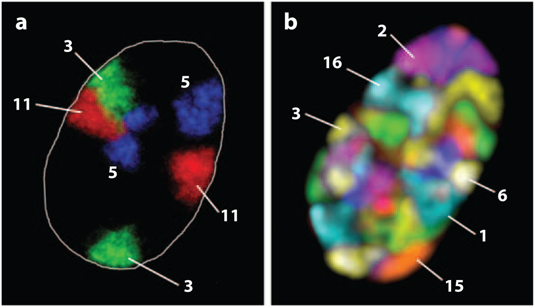
Figure 1.
Projections of mid-optical sections of human fibroblast nuclei that highlight chromosome territories. Three (a) and all 23 (b) pairs of chromosomes were detected using 3D-FISH with chromosome paint probes obtained by flow-sorting. Individual chromosomes are indicated. Image in panel a courtesy of Irina Solovei. Image in panel b courtesy of Andreas Bolzer and Irina Solovei, University of Munich, Germany.

