
Fig. 1.
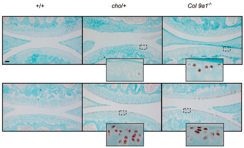
Immunostaining for HtrA1 of articular cartilage taken from the knee joints of chol+, Col9a1−/− and wild-type mice. Immunostgaining intensity of HtrA1 (see brown color staining) was increased in focal areas of the articular cartilage of Col9a1−/− mice, at 3 and 6 months of age, and of cho/+ mice, at 6 months of age, when compared to that of wild-type mice. The top panel reprsents samples taken from 3-month old mice and the bottom panel represents samples taken from 6-months old mice. The presence of positively staining cells in the 3-month old of Col9a1−/− mice and the absence of such cells in the 3-month old cho/+ mice suggests that the degenerative process in Col9a1−/− mice is much more advanced than it is in cho/− mice. This is consistent with the morphological changes observed in the articular cartilage from the knee joints of cho/+ and Col9a1−/− mice. Bar: 50 μm.