
Fig. 2.
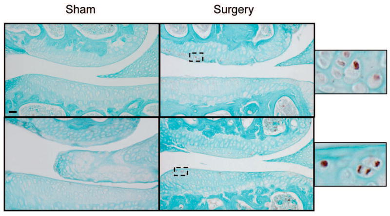
Immunostaining for HtrA1 of articular cartilage taken from surgically-induced OA knee joints. The onset of articular cartilage degeneration in the knee joints of surgically-induced mice was observed at 4 weeks post-surgery. At that point in time, focal areas of the knee joints of surgically-induced mice exhibited increased levels of immunostaining for HtrA1 (see brown color staining), when compared with those of sham-surgical mice (top panel). This relative increase in immunostaining intensity for HtrA1 was also evident in the articular cartilage of surgically-induced mouse knee joints at 8 weeks (bottom panel) post-surgery. Bar: 50 μm.