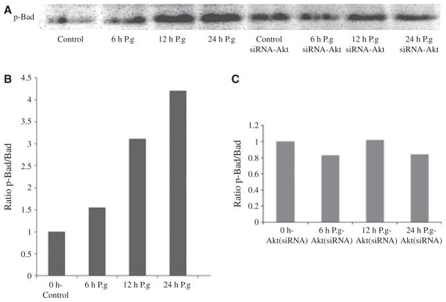
Figure 3.
Porphyromonas gingivalis infection induces a large gradual increase in Bad phosphorylation. Primary gingival epithelial cells (GECs) and the Akt-deficient GECs were infected with P. gingivalis for 0 min (control), 6, 12, and 24 h. Cell lysates immunoprecipitated with anti-Bad-specific antibody analysed by immunoblotting with antibodies against phosphorylated Bad (Ser136) (A). Blots were analysed by scanning densitometry and ratios of phosphorylated:total Bad were determined relative to ratios in control cells. The values show relative fold change calculated for a representative experiment and represent results obtained from at least three experiments (B,C).

