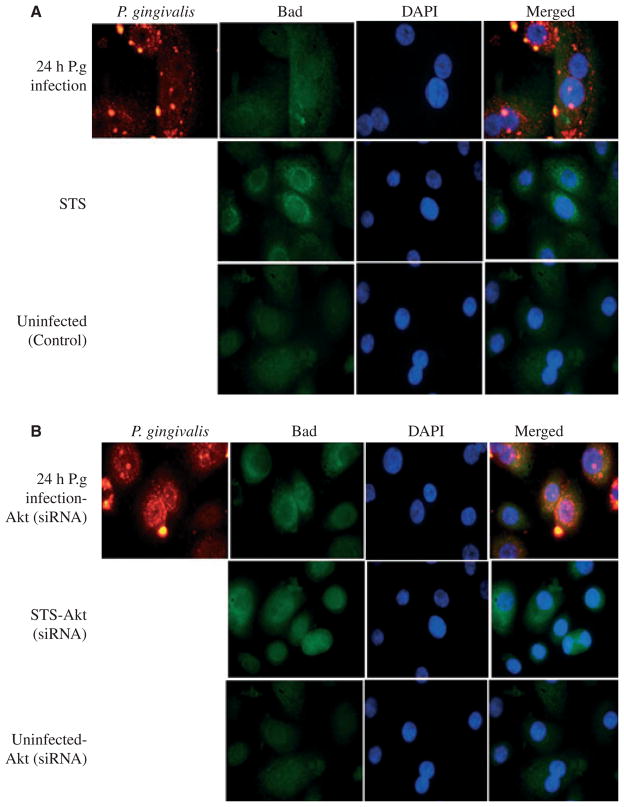
Figure 5.
Porphyromonas gingivalis infection redistributes Bad localization in primary gingival epithelial cells (GECs) through Akt. Intracellular Bad localization was detected by immunofluorescence using antibodies against Bad (green). The samples were also stained with P. gingivalis antibody (red) and DAPI (blue) to visualize the nuclei. (A) Uninfected cells displayed large proportions of Bad localized in the cytosol. However, incubation with the apoptosis inducer staurosporine (STS) caused strong staining in the perinuclear area, indicating translocation of Bad to mitochondria. Infection with P. gingivalis produced sequestration of Bad in the cytosol. (B) Akt knockdown cells were prepared as in (A). The localization of Bad showed intense staining around the nuclei, where the mitochondria are. This was similar in the uninfected STS-treated cells (control). The images were captured with a fluorescence microscope equipped with a cooled CCD.

