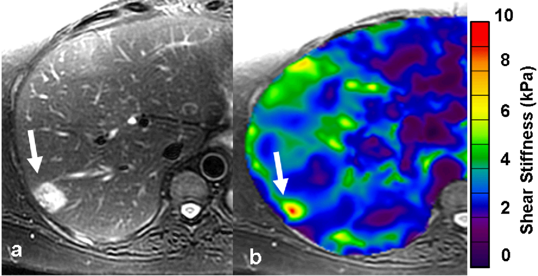
Fig. 4.
55-year-old female with fatty liver and a focal lesion in right lobe. T2-weighted image (a) showing a single hyperintense lesion in the periphery of right lobe of the liver (arrow). Elastogram shows the tumor as a “hot spot” with stiffness value of 6.2kPa suggestive of a malignant tumor. A subsequent colonoscopy revealed a carcinoma in the recto-sigmoid region. Patient underwent surgical resection of the liver tumor and confirmed to be metastases.

