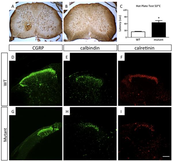
Figure 4. Anatomical and Nociceptive Defects in Hoxb8 Mutant Mice.
(A and B) Spinal cord sections at cervical levels in (A) wild-type mice and (B) Hoxb8 mutants stained with anti-NeuN antibody. These sections are representative of wild-type and Hoxb8 mutant sections taken along the spinal cord from C4 through the lumbar region L5. Neuron counts are decreased and the remaining interneurons noticeably disorganized in the mutant spinal laminae. (C) The latency of response to heat at 53°C, was significantly increased in Hoxb8 mutants. White bar, control siblings; black bar, Hoxb8 mutant mice. Columns represent the mean ± 1SEM. (D - K) Anatomical defects in dorsal spinal cord of Hoxb8 mutant mice. Spinal cord sections shown at L4-L5 from (D-F) wild-type mice and (G-I) Hoxb8 mutant mice. The spinal cord sections were labeled with a marker for nociceptive sensory fibers (CGRP), and with interneuron markers for lamina I and II (calbindin and calretinin). The number of interneurons in laminae I and II, are decreased and disorganized in Hoxb8 mutant mice relative to wild-type mice. Scale bar (D – I). 100μM. See supplemental Figure 4.

