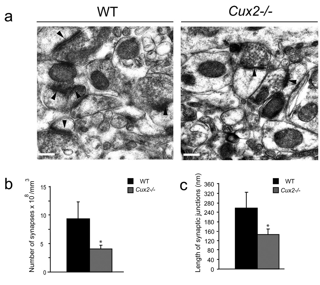
Figure 3. Altered synapse formation in the upper layers of Cux2−/− mice.
a)Electron micrographs showing the synapses (arrowheads) in sections of cortical layers II–III of the somatosensory cortex of WT and Cux2−/− animals. Bar represents 0.25 µm. b) Quantification of synapse density in layers II–III of WT and Cux2−/− animals. c) Average length of the synaptic junction apposition surface in layers II–III of WT and Cux2−/− animals. * p<0.001 compared with WT.

