
Figure 1.
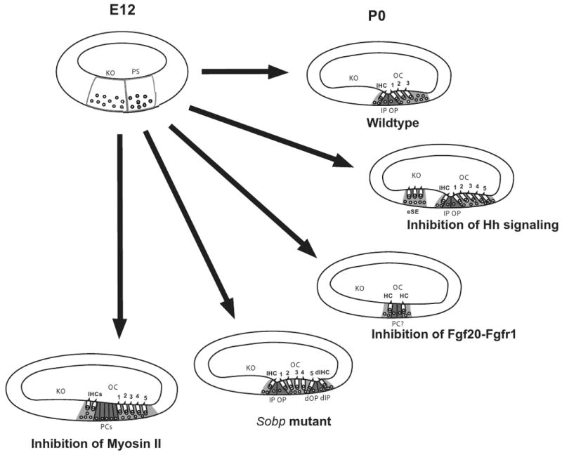
Phenotypic changes in the organ of Corti in response to changes in specific signaling pathways. At E12, the floor of the developing cochlear duct is comprised of undifferentiated cells that can be classified as part of the prosensory domain (PS) or Kolliker's organ (KO) based on relative position. By P0, the normal cellular pattern of the organ of Corti (OC), including a single inner hair cell (IHC), three outer hair cells (numbered), inner and outer pillar cells (IP and OP) and supporting cells (light gray), is present. Inhibition of hedgehog signaling leads to the formation of ectopic hair cells and supporting cells (eSE) in Kolliker's organ as well as an overproduction of outer hair cells. In contrast, inhibition of Fgf20-Fgfr1 interactions leads to the formation of small patches of unpatterned hair cells and supporting cells including some cells that may be pillar cells. In Sobp mutants, mirror-image duplications of the organ of Corti are present including a duplicated inner hair cells (dIHC) and duplicated pillar cells (dIP and dOP). Finally, inhibition of Myosin II leads to defects in extension of the cochlear duct. In a cross-section this appears as additional rows of all cell types.