
Figure 1. Direct assessment of BoNT/G binding to Syt and GT1b.
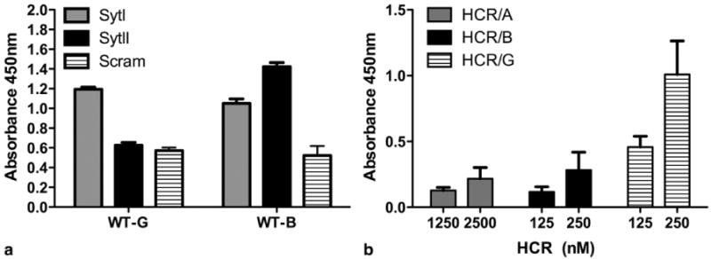
a) Peptides from the luminal domains of SytI or SytII (or a scrambled negative control) were covalently linked to individual wells of maleimide-activated 96-well plates through N-terminal cysteine residues. Unbound peptide was washed away, unreacted maleimide groups were quenched, and wells were incubated with excess BoNT/G- and BoNT/B- HCR (25 μg/ml) for 2 hours at room temperature. Wells were washed 5 times and incubated with His-Probe (an NTA-horse radish peroxidase fusion). Addition of a TMB-ELISA substrate allowed colorimetric detection of bound protein. The average (with standard deviation) for five replicates is shown. b) Non-binding ELISA plates were coated with 10 μg of GT1b / well in methanol and dried overnight. Plates were blocked in carbonate buffer, washed 3× with PBS and incubated with the indicated HCR-Hisprobe conjugate for 1 hr at 4°C. The plate was washed and developed with TMB substrate for 30 min, stopped with 1M H2SO4, and absorbance read at 450nm. Duplicate wells were averaged and subtracted from absorbance detected in an uncoated well. Data are the average of two independent experiments. In both experiments, the mutated form of BoNT/G containing the EERYK solubility sequence was used. The 6xHis sequences are located at the N-terminus of the HCR domains and are not expected to interfere with Syt or ganglioside binding.