
Figure 3. Structural conservation in the Syt binding site.
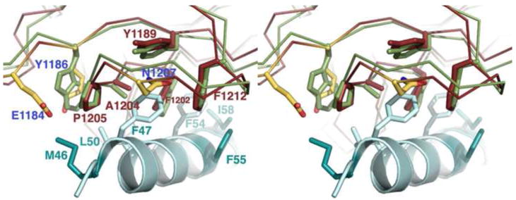
A cross-eye stereo close-up of the F47 binding pocket indicates that five of the 12 residues making contact between BoNT/B and SytII are conserved in both identity and position in the structure of BoNT/G (shown as red or green sticks to represent BoNT/G or BoNT/B, respectively). A sixth contact residue (BoNT/B W1178) is replaced by Y1186 in the BoNT/G structure. BoNT/G residues E1184 and N1207 (colored by atom) do not align with BoNT/B contact residues but are positioned so that they could impact Syt binding. The coordinates for BoNT/B were taken from the 1EPW structure while the SytII peptide (teal) is taken from the aligned 2NM1 structure. Residues that differ between SytI and SytII are shown in dark teal while conserved BoNT/B contact residues are shown in light teal.