Fig. 5.
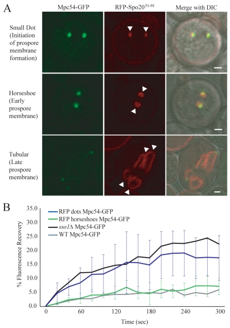
Vesicle fusion into a membrane structure reduces the exchange of MOP components. (A) Stages of prospore-membrane formation. Images are from wild-type cells coexpressing Mpc54p-GFP and RFP-Spo20p51-91. Arrowheads indicate prospore membranes. Scale bars: 1 μm. (B) Fluorescence recovery of Mpc54p-GFP from cells with RFP dots and from cells with RFP during meiosis II. The plots represent the average of nine and eight experiments, respectively. Error bars represent the s.d. at each time point. Fluorescence recovery of Mpc54p-GFP wild-type and sso1Δ cells from Fig. 1D are also shown for comparison.

