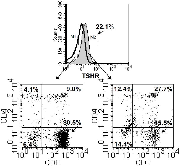
Fig. 5.
Three-color staining of small intestinal IELs for expression of TSHR, CD4, and CD8. Freshly-isolated IELs from normal mice were reacted with biotinylated bovine TSH and streptavidin-APC, plus PE-labeled anti-CD4 and FITC-labeled anti-CD8 mAbs as described in the Materials and methods. Control staining consisted of cells treated with streptavidin-APC, plus PE-labeled anti-CD4 and FITC-labeled anti-CD8 mAbs. In this experiment, the TSHR was expressed on 22.1% of the total IELs. The IELs consisted of four cell populations based on CD4 and CD8 expression, in proportions typical of IELs from normal mice [24]. Although the TSHR was expressed on all four cell populations, the majority of the TSHR+ IELs were CD8+ cells that included CD4−CD8+ and CD4+CD8+ cells. The remaining TSHR+ cells were CD4+CD8− cells or CD8−CD4− cells. These findings demonstrate that some but not all IELs are TSH-responsive cells, and they indicate that TSH utilization is focused within specific groups of intestinal T cells. Results are representative of experiments from three mice.

