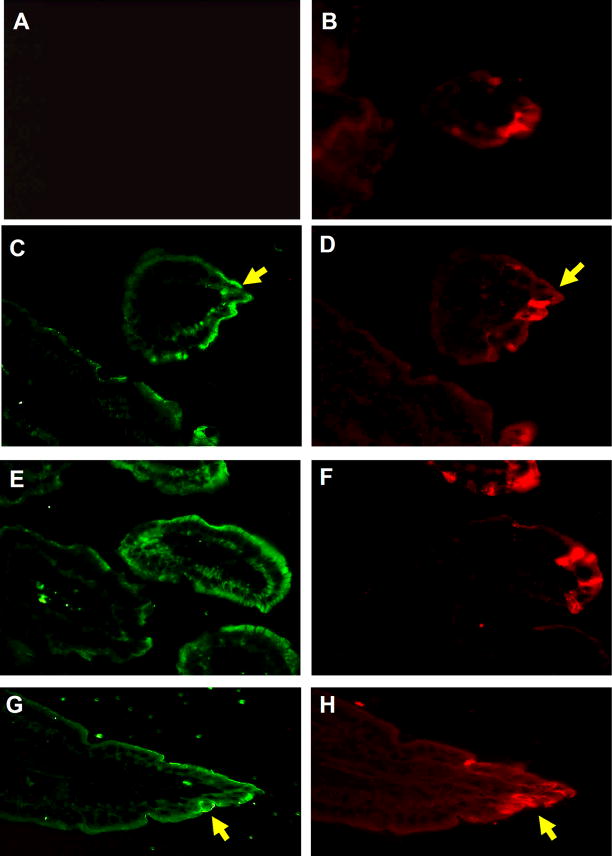
Fig. 7.
TSH and rotavirus staining of intestinal epithelia at 2 and 3 days post-infection. Staining of intestinal epithelia from mice 2 days post-infection (panel C and D) and 3 days post-infection (panel E–H) reveals TSH and rotavirus staining in villus tips the small jejunum. Panel A: staining of intestinal tissues with biotin-labeled control Ig plus streptavidin-FITC from (panel B) a rotavirus-infected mouse indicates that TSH staining of tissues was not due to non-specific antibody reactivity in infected regions. Results are representative of three infected mice. Magnifications: Panel A–H, 400x. Yellow arrows highlight some, though not all, of the regions where TSH staining and rotavirus staining are common.

